Ataköy 7-8-9-10 Ataköy Towers A Blok Kat 14 No: 169, 34156 Bakırköy/İstanbul
Microscopes and magnification in dentistry: What are they for and when are they used?
Microscopes and magnification in dentistry: What are they for and when are they used?
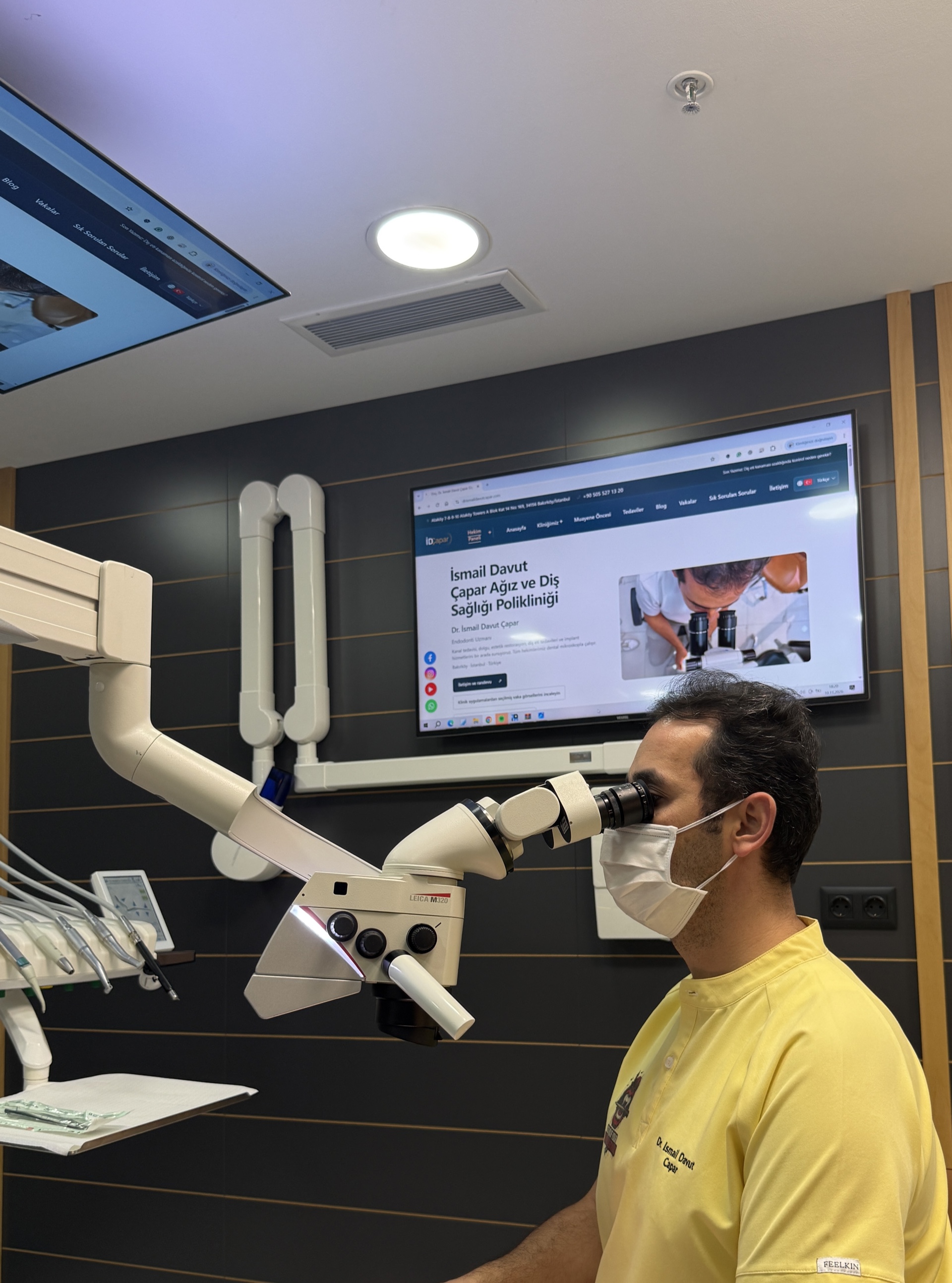
Some parts of teeth and root canals are very small. Dentists may use magnifying glasses or a dental microscope when assessing these areas. Magnification makes details in the field of view easier to distinguish; the microscope's light also helps illuminate the working area. These tools support the dentist's examination and treatment planning.
What is the difference between a dental microscope and magnifying glasses?
Magnifying glasses, also called dental loupes, are optical aids attached to or worn like glasses. A dental microscope is positioned near the dental chair and provides adjustable magnification and lighting. It can help the dentist examine a smaller part of the field of view in detail. The appropriate tool depends on the procedure and working conditions. Greater magnification does not automatically mean a better result for every procedure.
How can a microscope help during root canal treatment?
In root canal treatment, the canals inside the tooth are cleaned, shaped and filled. Their number, shape and width can vary from tooth to tooth. Magnification and lighting can help the dentist particularly when:
- Looking for canal openings and assessing the tooth's internal anatomy.
- Seeing the working area in more detail while examining narrowed or calcified areas.
- Assessing existing filling material or obstacles inside the canals of a previously treated tooth.
- Seeing a broken instrument in a canal and assessing whether it can be removed.
- Examining certain crack lines or unintended openings in the tooth.
The procedure differs in each situation. For example, a broken instrument cannot always be removed; its position, the structure of the root and possible risks are assessed before discussing the options.
Can magnification be used for fillings and other procedures?
Yes. Magnification can be used to assess areas of decay or fracture, examine the edges of fillings and other restorations, and perform some gum or surgical procedures. The appropriate tool and level of magnification depend on the procedure. Research suggests that magnification can support visual assessment in some procedures, but the evidence is insufficient to say that higher magnification is better than lower magnification in every procedure.
Does a microscope replace an X-ray?
No. A microscope magnifies areas that the dentist can see directly; it does not show every area inside the tooth or surrounding tissues. X-rays provide different information. The dentist considers symptoms and examination findings to decide whether imaging is needed and which method is appropriate.
What does the patient feel during treatment?
The microscope is not placed in the patient's mouth; the dentist looks at the treatment area through the device. The light may be directed towards the patient's face or mouth. Tell your dentist if the light is uncomfortable, if you find it difficult to keep your mouth open, or if you experience another problem. Pain control and other measures are planned according to the procedure and the person's circumstances when needed.
Is treatment with a microscope always successful?
No. Although magnification and lighting help reveal details, the outcome depends on many factors, including the condition of the tooth and surrounding tissues, canal anatomy, infection, the remaining healthy tooth structure, the treatment performed and the restoration placed afterwards. Studies have examined the contribution of magnification in some areas; however, evidence that establishes its effect on long-term outcomes independently is limited, particularly in non-surgical root canal treatment.
Is a microscope necessary for every dental treatment?
The same magnification tool is not necessary for every procedure. After examining you, your dentist can explain the condition of the tooth, treatment options and tools that may be used. You can ask about the aim of treatment, its possible limitations and the need for follow-up.
This article is for general information. Diagnosis and treatment planning are based on an individual examination and any assessments considered necessary.
Sources
- American Association of Endodontists. The Dental Operating Microscope in Endodontics. 2016. https://www.aae.org/specialty/wp-content/uploads/sites/2/2023/12/Winter2016-DentalOperatingMicroscopes.pdf
- American Association of Endodontists. Treatment Standards: Standards in Access Cavity Preparation. 2019. https://www.aae.org/specialty/wp-content/uploads/sites/2/2019/11/TreatmentStandards_2019.pdf
- Del Fabbro M, Taschieri S. Endodontic therapy using magnification devices: a systematic review. J Dent. 2010;38:269–275. https://pubmed.ncbi.nlm.nih.gov/20117164/
- Ucmak S. The Dental Operating Microscope in Endodontics: A Narrative Review of Clinical Applications, Supporting Evidence, and Limitations. Cureus. 2026;18:e117039. https://pubmed.ncbi.nlm.nih.gov/42807697/
- Effects of magnification on restorative dental preparation performance: a scoping review and level of evidence mapping. https://pubmed.ncbi.nlm.nih.gov/39052037/
Website editor contact: drismailcapar@gmail.com
